Sunday 2 September 2012

Introduction to Integumentary System

Hello Readers!!!!!!!

Today, I will write an introduction article to the Integumentary System of our body. Integumentary System is protective layer of our body, i.e., the skin, the Integumentary System consists of the Skin. It account for about 6% of the total body weight.


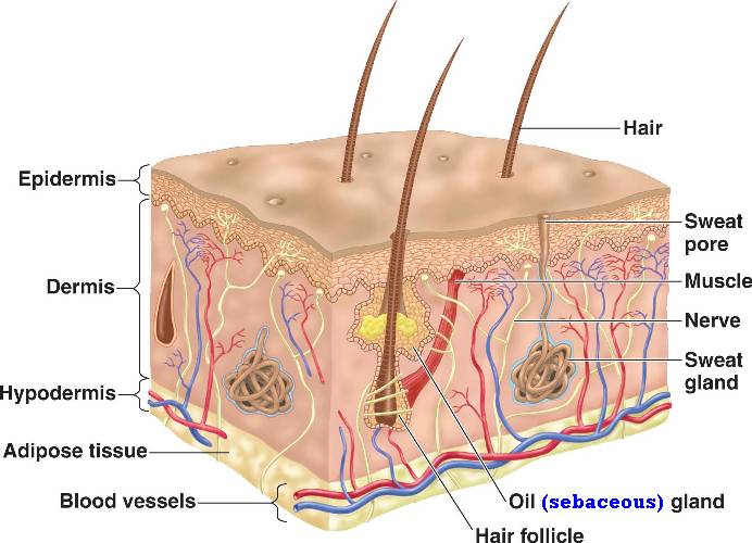
The above diagram shows a brief view of the Integumentary System. I will tell you about some of the cells in the Integumentary System and also the three main parts of the body. The skin is mainly divided into three parts:

  1. Epidermis.
  2. Dermis.
  3. Subcutaneous Layer or Hyperdermis.
Epidermis is the most superficial layer of the human skin which protects us and separates the internal environment of the body from the external environment.


The above image shows the Epidermis. The Epidermis is like the army of the skin which defends the boundaries of the human body. As this article is a brief discussion on the Epidermis, I will just give you a review over the parts of Epidermis. The Epidermis is made up of different cells named Keratinocytes, Melanocytes, Merkel Cell, and Langerhans Cells. The keratinocytes are the cells that make up a protein named Keratin, which is an important protein which provides strength to the skin. Next comes the Melanocytes, these cells are the cells that secrete a substance named Melanin, Melanin is a substance which gives your skin the color and this melanin also protects the Nucleus of the Keratinocytes from the Ultra Violet (UV) Rays of the sun. This UV rays can damage the DNA of the nucleus of the cells as it changes the genetic information in them. But the Melanocytes are sometimes mostly themselves damaged due to UV light.

The Merkel Cells are the cells that are joined to the nerve endings of the neurons that are connected at the base of the Epidermis. The Merkel Cells are sensitive to touch and without them, we cannot feel the touch of any person or any other thing.

The Langerhans Cells work like the Police in the Epidermis as they prevent any Microbial Invasions in our body by secreting several antibodies which fight against them. This Cells actually arise from the B Lymphocyte in the bone and then travel to the Epidermis in order to protect it from infections by microbes.

There are also several layers of the Epidermis namely Stratum Basale, Stratum Spinosum, Stratum Granulosum, Stratum Luicidum, Stratum Corneum. The Stratum Basale comes at the last and the Stratum Corneum is superficial, i.e., it is located at the outermost part of the skin.The below picture shows some information about it:


The Stratum Corneum which is the superficial layer contains dead Keratinocytes which are not at all functional now, they die because the supply of blood vessels is more at the Basale layer and as the cells go away from their nutrient supply that is the blood vessels, they eventually die. In the Stratum Granulosum, the Keratinocytes start producing Lamellar granules which provide strength to the Layers of the Epidermis.

Now let's talk about the Dermis, the Dermis has two layer namely Papillary region and the reticular region.

The papillary region is the region that is the superficial layer of the dermis and this part contains the dermal papillae. The dermal papillae is shown in the below image:


The dermal Papillae bulges out as shown in the above diagram an the bulges and just underneath the Epidermis. This increases the surface area and thus allowing the Epidermis to fix very well with the Dermis. The number of dermal papillae varies from place to place. They also contain the Capillaries which provide the blood to the Epidermis. It also has the Meissner Corpuscles, this are the nerve ending located in the Dermis that are sensitive to touch. The Papillary region can also be thick in some regions and can be thin in some areas.

The next layer is the Reticular region which is just located inferior(below) the Papillary region. The reticular region anchors almost all types of things such as hair etc. that arise from it. This layer of the dermis has a net like structure of the fibres in it and in between the spaces in the net like formation, the hair and other things have their roots.

Now, let's talk about the Subcutaneous Layer or it is also known as Hypodermis. This layer is the layer that contains some oil glands and also some of the adipose tissues, i.e., the tissues that contain the fats. It also serves as the protector of the bone and the muscle from the shocks as the adipose tissue absorbs it. It also has the lamellated corpuscles that are the nerve endings that are sensitive to the pressure.

This three layer are the layers that constitute the Integumentary System.

Thanks for reading this article and sorry for any type of mistakes I made in the article.
Comments are appreciated.
Have a good day!!!!!!!!!!!

Sunday 26 August 2012

Neil Armstong

The day of 25 August,2012 was a very sad day for the whole humanity because one of the World's Greatest Astronaut Neil Armstrong died because of some cardiovascular problems. He was the first person to pass the greatest leap of humanity by stepping on the moon. He took the humanity out of this Earth and made it realize that it is had not discovered everything. If you really want to salute that person, who made history in humanity, then please share this article.

I wished to post some photos of the great person but unfortunately because of copyright, I am not able to do so. But I really salute that person.

Thanks for reading and please share it or give it a G+ if you want to salute Neil Armstrong. 

Saturday 18 August 2012

Cell anatomy videos.

Hello my dear readers!!!!!!!!

I know that sometimes we don't have time to read much of the things that I write, so I came up with a new idea of sharing some of the great videos on cell anatomy which will surely make you understand them very carefully. So, here are some of the videos, enjoy them, understand them, and also save you time.


Hope you enjoy this videos, next I will post videos about Tissue Anatomy.

Enjoy them.

Wednesday 15 August 2012

Alzheimer's-type dementia

Today I will be talking about Alzheimer's-type dementia which is a neurological disease. But before I start, I want to tell you all about the correct pronunciation of the word neuron. It is pronounced as NOO-RONS. I wanted to tell this thing many people including me thought that it was pronounced as NEW-RONS. Now let's get back to the main topic.

Dementia is a neurological disorder in which people usually suffer from certain symptoms such as they are not able to memorize thing's, for example, they might buy on loaf of bread two times from the same shop even if they require one as they won't remember that they had got one already. A dementia can effect the person suffering from it in various ways. It can cause the person to behave in an aggressive way and also can have effect on their language. As a result, they are not able to pronounce anything clearly and they might forget the pronunciation of a word. It also affect's their mental abilities such as doing calculation's and also their motor functions. The motor functions means the movement. As a result of this, they might not be able to do even the easiest things, such as brushing the teeth, eating food, taking bath or driving.

The Dementia is classified into several types, on of the main dementia is Alzheimer's-type Dementia or simply Alzheimer's Disease. In this disease, the brain cell's gradually start's depleting and the ventricles in our brain(the hollow spaces in our brain) starts increasing in size, and because of this, the brain's size starts decreasing and at an stage of severe Alzheimer's disease, the person dies because most of the parts of his or her brain have stopped working.

We usually detect if a person was suffering from Alzheimer's Disease after his death while autopsy. And sadly, their are currently no better cure for this type of disease. The scientists have divided the Alzheimer's Disease into ten stages named S1 to S10. We usually see the effects of this disease or we usually recognize that the patient is suffering from this disease after he had reached the stage of S6. Alzheimer's Disease is really very harmful disease and it is also not good that it is not having any type of cure right not and we are on the search for finding cure.

Thanks for reading and I think this much information is adequate for you right now.

Friday 27 July 2012

Windows 8 COMING SOON!!!!

Hello friends!!!!!!!!!!!

Today I have a great news for the people who are a type of computer geek. Microsoft had announced that soon they will release the Windows 8 version for the public to buy on October 26, 2012. Microsoft had already released the Windows 8 Developer and Release Preview. If you want to feel the windows 8 before it is commercially launched, then you can download the Windows 8 Release Preview from the link below:

http://windows.microsoft.com/en-US/windows-8/download

But remember to use dual boot setting to run windows 8 with windows 7 as it is said that Windows 8 Release Preview can be buggy and can stop working suddenly. Below is the link from where you can learn how to do dual boot:

http://www.howtogeek.com/107535/how-to-seamlessly-dual-boot-windows-7-and-windows-8-the-easy-way/

But is don't like the Windows 8 Release Preview, then you might require to wait till October 26, 2012. But don't worry, because Microsoft had also announced that it will be releasing the Windows 8 RTM version which would be just like the commercial version for the Manufacturer's of Windows 8. So if you have some friend or relative working in the Microsoft then you can ask him to give you a look at it or just install it on your computer.

Thank's for reading my friend's!!!!!!!!!!!!!!


Saturday 21 July 2012

Types of stroke.

Hello my dear reader's!!!!!!!

Today Stanford will tell you about the types of strokes in our brain. Just watch the video below:


Here are the notes that you can revise after watching the video:


Neurons communicate through Ions. Ions are passed through the cell membrane and the inside of the cell is negatively charged while the outside is positively charged. The neurons send electric signals to communicate known as action potential. The difference between the cell's inside and outside atmosphere is maintained by the proteins in the cell membrane.
They do so by pumping certain ions inside and certain ions outside,this pumping actino requires energy.
Neurons only work in aerobic conditions and they don't have their own energy stores. Brain only costs 2% of body weight, but requires about 20% of cardiac output, and 15% of oxygen take from the lungs. The blood to the brain reaches from the aorta is taken up to the brain by the carotid artery,
which takes 70% of it. The other 30% is taken up by basilar artery and verebral artery. This two artery are joined the base
of the skull and are then divided into six parts (2 parts each) -

1. Anterior Cerebral Artery.
2. Middle Cerebral Artery.
3. Posterior Cerebral Artery.

The Circle of Willis is the place where all these arteries join to provide the brain with blood. This circle joined the right and left artries so that if one artery (either left or right) is not working, then the brain will still receive the blood instead of no blood. Now there are also many other deeper parts in our brain, this parts will get supplied by different arteries. If this arteries don't work properly, that certain part might not be able to function properly.
For example, if the blood does not reach the Broca's and Wernickel's area, this parts will stop working and that person might suffer from Broca's Affesia, a disease in which that person will have language related problems.

A stroke is defined to be an abrupt onset of focal or global neurological symptoms, caused by ischemia or hemorrhage.
Ischemia means restriction of blood in Greek. And it is usually caused due to factors in blood vessels, with resultant damage in tissues. Tissue damage is caused by shortage of O2 or Glucose.

Hamorrhage is discharge of blood from blood vessels AKA bleeding. Bleeding in the tissue or around the brain. Their are 3 types of strokes, Ischemic stroke, Intracerebral Hemorrhage, Subarachnoid Hemorrhage. Ischemic Strokes are usually caused by blood clotting in the brain which will result to no O2 and glucose supply to the brain. Intracerebral Hemorrhage is bleeding in the tissues and Subarachnold Hamorrhage is bleeding around the brain.

Warning signs of Stroke:

1. Sudden Numbness on the face or any other body part, especially on the one side of body.
2. Sudden Problems in speaking or understanding.
3. Sudden, severe headache with no known cause.
4. Sudden trouble walking, loss of balance or coordination, dizziness.
5. Sudden trouble seeing with one or both of our eyes.

If a patient walks in and says he/she has a stroke, we can do the image studying of the brain to confirm that, the images show us what type of stroke it is or either it has an other cause such as tumor, infection, epilepsy. Imaging also tells us how better the brain is getting blood from the blood vessels. Their are two methods of Imaging, MRI an CT scan.

We can also use the cerebral angiography scan to determine the stroke. The main symptomes of strokes are usually seen in people with High blood pressure, diabetes, people who smoke, drink or intake tobacco.

Ischemic stroke is caused when the thrombus is formed which leads to vessel occulsion leading to O2 depletion leads to membrane ion failure which leads to cell swelling and then cell death. Some thrombi take time to form but there are some other thrombi that are formed in the heart and are then sent to the brain from the heart. Some strokes can also be temporary but we must remember that they may be a sign of a stroke. tPA is a method to diagnose the deficits of stroke, but it is not very much effective. The occulsion of a cerebral vessel is followed by formation of a central irreversible infarct, with a peripheral zone, the penumbra, where the tissue is variable due to partial preservation of blood supply via collateral vessels. CT Perfusion shows up the penumbra and infracted area. CT Scanning uses the X-Rays to make a
photo of the brain, the X-Rays are projected to the patient by the X-Ray generator which rotates around the patient to get a good view of the brain, the image of the brain is regerated by a process known as Back Projection as the X-Rays are changed in velocity when they interact with the tissues. MRI works with the help of the photon molecules or molecules containing photons, the MRI take advantage of the photon as they spin at a certain frequency at certain chemical environment, so we use different photons for different tissues, the photons when are bought in an magnetic field, then releases some enery as protons which we can record as they pass through the tissue to get an image of the brain. Rcently, Stanford University and other Universities have made a device that can go through the artery and the surgeon will pass the
device until the surgeon see's the thrombus, afterward's, the device will pull out a string through the clot, and will then turn the string into spiral shape so that the thrombus is stuck in it without any problem and will then just drag the thrombus with the help of the string and take it out. Another thrombactic device is also there that can relatively suck the clot in it and then the device can be taken out of the artery. If the thrombus is sent through the heart, after it is removed, there are sligth chances that the heart might send another thrombus again, which would again cause the ischemia. To make sure that this does not happen, the patient is passed through certain medication after the thrombus is removed from the artery.
The cerebrovascular system is the only route for energy supply to the brain. Blood vessels are lining organs that are under constant state of vascular remodeling. Acquired and cognital problems of this system are important causes of stroke.
In hamorrhage, the blood vessels spell the blood in or around the brain, and as a result of that, that particular vessel does not supply that particular brain part with which it is associated and this leads to ischemic stroke. More people die because of hamorrhagic stroke than the ischemic stroke. The causes of hamorrhage are divided into Primary and Secondary causes, primary cause of hamorrhage is hypertension etc. Ischemic stroke can also be converted to hamorrhage as in
ischemic stroke the blood is not able to reach in the brain which can make the vessel blast as the thrombus won't allow the blood to pass through the artery and then the artery will not be able to take on the blood pressure and will blast off.
Ischemia can also lead to hypertension which would lead to hamorrhage. Surgery is the best way to treat hamorrhage but there are chances of accident in surgery which can lead to brain damage. Aneurysms are the defect in blood vessels where the blister's form. We don't know why the aneurysms occur. The people who suffer from aneurysms have brother's and sister's who also suffer from this aneurysms, this makes us think that there is some genetic problems related with it. The larger the aneurysms the bigger the chance to blow. There are several causes of SAH (Subarachnoid Hamorrhage) such as hypertension, alcohol, pregnancy, drug or cocaine, smoking.
 

Aneurysms usually happen where two arteries join. Aneurysms can be found in a Stroke, Seizure, or a headache pain.
We can tell if a patient has SAH if he is reporting a very worst headache of his/her life, nausia, vomiting, photophobia, neck pain, less control over eye or face, altered level of consciousness, sential headaches (warning leaks) may proceed major, clinically divastating SAH as in half of the cases. First thing to diagnose a SAH is to get an angiographic study of it. We can also do a 3D angiogram. We can treat aneurysms by surgery and enduvascular coiling. In the surgery, we first make a small window in the scalp to look inside the brain, and then we look through a microscope in it, find the aneurysms and then tie a clip around the aneurysms, this would not allow the blood to get into the aneurysms and so the aneurysms
will not bleed out. In endovascular coiling, we tie the aneurysms from the bottom to the top so that the blood can't enter into the aneurysms. The coil comes of different types and sizes. The endovascular coiling can be done with the Primary treatment, Ballon assisted treatment, and Stent assisted treatment. In primary treatment where the neck is not to much large, we just coil the aneurysms, but in ballon assisted aneurysms, we need to use a ballon so that the coil did not fall back to the vessel. There is a device known as flow diverter that can divert the flow of the blood into other blood vessel so that the surgeon can perform the surgery. Arteriovenous Malformations is an abnormal collection of blood vessels wherein
the arterial blood flow directly into the veins without proper capillary beds. Cognitial , but may enlarge with age, this may involve an abnormality or dysregulation of vascular development or remodelling. Usually in AVM's, they are seen like very large capillaries and the veins in AVM usually loss the two layers of their walls, as well as we are not able to see the brain tissue between them, this condition is named as enciephalomalacia. This may be due ischemia at that place or due to the hamorrhage as the veins are carrying blood at high pressure and they are not accustomed to it. This disease usually
is diagnosed at the adulthood as it starts creating problems at that time and it is a rare disease (1-2 people per 100,000)
We use CT (CT Angiography),MRI (MR Angiography), or angiogram to find AVM's. We can cure AVM's by using microneurosurgery, which is very good method for curing the less sophisticated AVM's as well as which are located on the upper side if the brain. AVM's might have been in the patient from their birth or it might have been developed with age but it usually is within the patients at their time of birth. The AVM usually occur when the capallaries are not able form the correct way and they grow thicker and thicker with the time. The AVM can also be treated with the help of Stereotactic Radiosurgery, in this process the AVM is cured with the help of radiation and this is only done when we usually cannot acces the AVM
surgically. We use Endovascular Embolization to make the microsurgey or the Stereotactic Radiosurgery more effective and this all is known as Multimodality approach,i.e., using two or three methods together to cure the Anteriovenous Malformation. The CyberKnife Radiosurgery is made by the Stanford Scientists and it is used to target the particular area directly by an image guided robot. Embolization helps us to get a 3d graph of the brain or the Anteriovenous Malformation as well as helping us to decide weather it is okay or not to do an surgery. We can also know about the risks of performing
a surgery there and also introduce flow-directed micro catheters to the patient's body so that will be assisting in the furture processes as well as to inject liquid in the blood which can literally block that portion of the blood vessel(AVM).
Galen AVM is a type of Anteriovenous Malformation in which the AVM is much dilated than the usual.

Thursday 5 July 2012

Sub - Woofer not working.

Hey guys!!!!!!!

Just few days ago, I was having a problem with my sub - woofer on my pc as it was not working. I tried to search on the google about this but I was not able to find any answers in it. They all said that it might be an problem in the wiring or an sound card defect. But I tried every thing and nothing works. But I got it working, here's how:


  1. First check if you have installed the right driver on your pc for the sound card or not. To check that, just see the speaker icon on the lower right corner of you moniter, if the speaker is with a red cross, this means that you have not installed the driver, install the correct driver or call the person from whom you bought the PC to know which drivers you need. Or if the speaker is with a stop sign with it, that means that you have accidentally turned off the volume. To turn it on, just drag the bar to the top.\
  2. (To perform this step, first close the PC and the UPS) If Step 1 does not work, then you might not be knowing that the sub - woofer also requires power to run which it usually gets from the UPS. Many people forget about it and buy a new Woofer. The thing you need to check here is that look at the back the sub - woofer and you will find a wire in it, other than the speaker wire or the CPU one, follow that wire and see at which socket is it installed, after you have turned the UPS off, then just remove the pin from that socket and replug it or try to use another working socket, this would give your woofer the power supply and you will be able to use it again.
  3. If the Step 2 don't work, then I recommend you to concern a professional in it.
Thanks for reading and I hope that your Sub - Woofer will work.
Comments are appreciated.


Wednesday 4 July 2012

Cell Division

Hello my fellow readers!!!!!!!!!!!!

Today I will be teaching you how a cell divides. First, I would like to tell you an amazing fact about cell division, our human body cells divides about a thousands of time in one minute. I found it very amazing and I think you will also find it also amazing.

Now let's get into the cell division process. First I need to tell you that the germ cells and somatic cells are the two classification of the cells in our body and these cells divide in different ways. Germ cells a.k.a. Gamete cells are the cells that are reproduction cells, such as ovum in women and sperm in men. And somatic cells are the cells that do not reproduce.

Their is one common phase in the Cell cycle that both the cell types undergo that is interphase. There are three phases in the interphase. They are:

  1. G1 Phase.
  2. S Phase.
  3. G2 Phase.
In the G1 Phase is the phase in which the cells start to divide. In this phase they just start the centrosome( An important part of cell) division and in this phase the cell is metabolically active and also the cell starts the division of cytosolic components.

During the S phase, the cell starts DNA replication, so that each type of new cell formed gets equal number of genes. Genes are a very important part of our cell, they are like the General Governor of the cell Army, each and every action usually starts from them. So, it is important that the genes must be divided equally so that the cell is able to perform the right way. The genes are the building units of the DNA.

During the G2 Phase, the centrosome replication is complete and the the DNA replication is also complete. At that time, the cell is usually synthesis proteins and other enzymes. But to be remembered that if some cell don't divide and the they are just locked in the G1 phase and are said to be in the G0 phase.

Interphase.


Now let's discuss about the mitosis and meiosis. The somatic cells divide through mitosis process and the germ cell divide through meiosis. First let's talk about mitosis, the mitosis is the process of cell division the includes 4 phases. Prophase, Metaphase, Anaphase, telophase.

In the Prophase, the cell starts dividing itself by first converting the Chromatids into Chromosomes. This is an important part as each cell should have the same amount of Chromosomes to perform the right way. Also in this stage, the cell's nuclear envelope breaks up and nucleoli disappears. The nucleoli is a part that is located within the nucleus of the cell. And the nuclear envelope protects the nucleus. And also in this phase the mitotic spindle starts to form. The mitotic Spindle is an important thing without which the cell could not divide.

To understand the process of mitosis better, here is an image :

Mitosis.

In the above image, we can see the full process of mitosis. In the image, the red and green marks indicate the chromosomes. The next phase is the Metaphase, the chromosomes are held together by centrosome, outside of the centrosome have a protein known as Kinetochore, the mitotic spindle attaches to them, and arrange them in a single line known as metaphasic plate. Then in the next phase, the chromosomes are separated into chromatids and they are pushed to the other side of the cells by the mitotic spindle. In this way, both the new cells will be having the same amount of chromatids. The next and the last phase in the process of mitosis is the telophase, in this phase,the nuclear envelope reappears and the parent cell is like pinched out in between to form the two new haploid cell.


The cytokinesis usually starts at the late anaphase and starts to divide the cytoplasmic material. It is usually completed in the telophase but the distribution is usually done roughly.

I will be writing the process of miesosis on later times because I think remembering this much today is still okay for you.

Thanks you for reading!!!!!!!!!!!


Thursday 14 June 2012

Limbic Lobe

As I have already talked about the basics parts of our brain, this part named Limbic Lobe is located inside the Cerebrum.

The upper diagram the shows the Limbic Lobe also known as Limbic System. In this article, I will talk about several functions of their parts.

The first part I would like to talk about is Amygdala. The Amygdala is a part in our brain that is mostly involved in processing emotions. Mostly the emotions of fear that alert us. For Example, An experiment was performed on a mouse,in that experiment, the researchers surgically stopped Amygdala from working. The researchers then putted the mouse in the cage of  a hungry cat, and they found that the mouse got into the hands of the cat very easily and the mouse was still not afraid, that is a sign that the Amygdala plays an important role in processing the emotion of fear.

Now let's talk about Hippocampus, Hippocampus is a part in our brain that is involved in making memory. I have already talked about it in my previous post and you can read that post for getting information about it.

Next I will talk about The Thalamas. The Thalamas is like a gateway for the Cortex, very information that wants to visit the Cortex must first pass through here to be able to go to the Cortex. The Cortex here might even cancel the information and it might not allow the information to reach the Cortex. For Example, when you are in your school and your teacher teaches you a very important lesson, you might not hear the tick tick sound of the clock, this is because the Thalamas after sometime does not allow the sound to reach the Cortex because the Cortex is already involved in understanding the lesson.

The Cingulate Gyrus is the part of our brain that processes the autonomic nervous system that is this part of our brain adjusts the Blood Pressure, Pupil Size, Heart Rate and other things which are not in our control.

Hypothalamas and Pituitary Gland.
The Hypothalamas is located just below the Thalamas and is mostly involved in maintaining the internal environment of our body by increasing or decreasing the feeling of Hunger, Thirst etc. It also affects the rate of our growth by getting a control over the Pituitary Gland. The Pituitary Gland secretes the Growth Hormone and it only sends the Growth Hormone when it gets the signal from Hypothalamas.

Next, I would like to talk about Corpus Callosum, This part of our brain helps both the hemispheres of our brain to contact with each other. There are two hemispheres of our brain, they are left and right hemisphere and this both contact each other with the help of Corpus Callosum.

The Limbic Lobe can be said as the heart of our brain. We cannot have emotions without it.

This is all I wanted to include in this article.

Thanks for reading.

Friday 1 June 2012

How electricity is generated?

Hello Friends!!!!

Today I will be going to give you some idea how a simple or we can say typical electric generator works.

First I would like to tell you what a electric generator is made up of, it is made up of 2 magnets, aluminium or copper wire and a turbine( or an object that rotates). The aluminium or copper wire are connected to the turbine and are placed between the 2 magnets. The magnets are such that magnet 1 would be placed with north pole facing the wire and the other will be placed in a position so that it's south pole will be facing the wire.

When the turbine starts to rotate, it also rotates the wire that is attached to it. When the wire placed between the magnets rotate, the repulsion force of the 2 magnets causes the electrons in the wire to move which creates electricity. You can also watch the video below to understand what I said.

Thanks for reading.

Saturday 12 May 2012

Basics Of Neuroscience

Hello Friends!!!!!!!
Today, in this article, I would be writing about the basis of Neuroscience.

First of all, I would like to tell you what is Neuroscience?

Neuroscience is a branch or part of science that  mostly deals with how our brain works. And this is one of the part of science that can tell you that your brain is not just a simple brain but it is really more complex than even a super computer. It's complexity is really hard to solve and study. So, in this article, I will only write about some of the basics of Neuroscience and also I recommend you to read my article which deals with the Neuromatrix of pain, i.e., how is pain caused and it tells about the parts of our brain involved in discovering pain.

So, let me first start with the basis of brain anatomy. Scientists have divided the brain into three parts in common. They are:

  1. Cerebellum
  2. Cerebrum
  3. And Medulla
First I would like to talk about Cerebrum. The Cerebrum is the part of our brain that almost processes all the data that comes in our brain through the spinal cord or all the information that comes into our brain. The image below shows the Cerebrum and it's four parts:
Human Brain

The cerebrum is again divided into four parts:
  1. Frontal Lobe
  2. Parietal Lobe
  3. Occipital Lobe
  4. And Temporal Lobe
First let me talk about the Frontal Lobe, this part of our(Human) brain is basically involved in planning and reasoning and all the other types of higher level functions that only human's can perform. More specifically, The Prefrontal Cortex is the part of our brain that is involved in higher order functions. And this part part, i.e., The Prefrontal Cortex, is only found in the human brain, that means that it is the special part that allowed us to perform the functions that animals cannot do.
Prefrontal Cortex
The image above shows the Prefrontal Cortex in green color. 

Now moving towards the other part of our brain, Parietal Lobe.

This part of our brain is mostly involved in the processing of movements. This part of our brain also helps us to keep an record of where our body parts are located in space, for example, close your eyes for some time and after you close your eyes, you still know where your body parts are or where you leg is right now, where you fingers are right now, so this is the part that makes that thing possible, or in other words, it contains a type of blueprint of all your body parts. 

This part also works in coordination with your occipital lobe which is involved in processing visual signals so that you are able to pick up a pencil when you see it.(discussed later)

As I have already said, that the Occipital Lobe is involved in processing visual signals. But for the people whoa re blind from birth may also use this part of their brain to process the auditory signals, it is usually found that the people that are blind from birth, have a greater sense of audio then any other normal person, the reason behind this is that this people usually train their Occipital Lobe to process audio.

Next comes our Temporal Lobe, the Temporal Lobe is a part in our brain which is involved in processing audio signals. It also contains the part of our brain, known as Hippocampus(HI-PO-KAM-PUS), that is involved in processing the memory portion, that is, the thing that we need to remember are processed here and are then sent to long term memory. It is like the gateway of the memory to be remembered. But one thing should be noticed that it only makes us remember the things but it does not store them, it sends that "thing" to the respective Lobe or Cortex it belongs to. For Example, the memory of audio will be sent to the Temporal Lobe, the memory of vision will be sent to the Occipital Lobe etc. Afterwards, this all the memories can be combined to form a image of what happened in the past or the thing we need to remember.
Hippocampus.

The Occipital Lobe also sends the signals to the Temporal Lobe and Parietal Lobe, for recognizing and picking up an object. The Temporal Lobe recognizes the object and the Parietal Lobe plans our actions to pick up the object.

Now we come to the part of Cerebellum, the Cerebellum is usually involved in coordinating motor functions, i.e., the functions of movement. The movement signal after getting processed in the Parietal Lobe, it reaches the Cerebellum. The Cerebellum also controls reflex functions. For Example, if you try to hit a trained person in self defense techniques, such as Bear Grylls, you will find that they have taken the decision to attack you in the correct manner in less than a second, the reason behind that is that the signal did not go to the Cerebrum, instead the Cerebellum used the neural connections in it to perform that action. And also you can modify the neural connections in the Cerebellum by practicing regularly.

Last comes our Spinal Cord, it is a part that takes signals from our brain to the body and also from the body to our brain.

I cannot write more than that right now as I am tired, sorry for any mistakes in this article.

Thanks for reading!!!!!!!!!

Friday 11 May 2012

WE DON'T HAVE WEIGHT!!!!!!!!!!

Hello Friends!!!!!

Today's topic is somewhat different because it says that we don't have any type of weight. This concept was thought by me and I think that we don't have any type of weight.

You might ask how can I say that? The answer is that you all know that we have a force called Gravity on Earth and this thing (Gravity) is also located in the moon. But as you all know that when a person on Earth goes to Moon, that person experiences some weight loss. But what is the reason behind this weight loss? The answer is that it is because there is less amount of gravity on moon then on earth. This leads to the conclusion that the Gravity has an effect on the weight of an object,this surely clears out that an if an object is sent in zero Gravity,then that object would experience zero weight. And also if any object is sent from a lower Gravity area to a Higher Gravity area, then that object's mass would increase.

This makes clear that we actually don't have any type of weight with us, it is just a part of Gravity, nothing other than that.

And I think that we don't have weight in zero gravity, then many formulae based on the weight need to be changed in Physics. For Example, The formula of Force is Force=Acceleration*Weight. And this formula cannot be applied in Zero Gravity because according to me, we don't have weight in Zero Gravity, so the formula may be changed to Force=Acceleration. Not only this formula, there are many other formulae that need to be changed in order to be applicable in Zero Gravity.

This also makes clear that the rules of Physics are not same everywhere, they cannot be same on Earth and a place with zero Gravity.

And that thing makes this research a noble one.

Thanks for reading.
Comments are appreciated.

Copyright© 2012 All rights reserved

Thursday 10 May 2012

Dentist recommend Ice Cream????????????

Hello Friends!!!!!!

Have you ever wondered that why the dentists recommend Ice Cream after removing a tooth????????

I wondered on this question and did some research on my is that so and I got the answer and I really think that it should be published on my blog so the other people also know that because curiosity is the best quality for any child or scientist.

Dentists recommend ice cream after taking out a tooth to just stop bleeding in the mouth and it really works but they don't say that we should always eat that because it might also cause cavities in our mouth, so we should eat it only one time after the dentist had taken out the tooth or till the bleeding does not stop.

We can also rake other cold things such as Cold Water.

Have a knowledge full day ahead!!!!!!

And thanks for reading this short article!!!!!!!!!!!  

Saturday 5 May 2012

VACATIONS!!!!!!!!!!!!!

Hello again guys and today I am happy to say that my vacations are going to start on 3 or 4 days and in the vacations I will not be able to post much articles but try my best to give atleast 30 minutes every day to write an article on this blog.

Thanks for your support.

Thursday 3 May 2012

Basic Anatomy of a Cell

Today I will be writing about the basic anatomy of the cell. The word anatomy here means the basic structure of the cell.
A cell.
Anatomy of a Cell.
The above image shows all the parts of a typical cell but I will not go in depth in this article about the structure of a Cell and will only write about some of it's part.

For the ease of study, the cell is divided into 3 parts:

  1. The innermost part:Nucleus.
  2. The part between plasma membrane and nucleus is Cytoplasm which contains mostly the organells(discussed later) and liquid.
  3. The outermost part is a protective layer of the membrane known as the Plasma Membrane.
First,I would like to discuss about the Plasma Membrane. The Plasma Membrane is the membrane the protects the inner parts of the cells and also regulates the particles that comes inside and goes outside the cell. Or in other words, compare this membrane to the hole in a balloon, the air can come inside and go outside only through that hole and the Plasma Membrane works the same way, they regulate the particles coming in and going out.

The Cytoplasm is the part that comes between the Plasma Membrane and the Nucleus. The Cytoplasm is divided into two parts:
  1. The Cytosol.
  2. The Organelles.
The Cytosol is the liquid which contains the organelles and organelles are the functioning unit of the cell.

Now moving towards the Nucleus. The nucleus is the part of our cell that is located at the innermost part of the cell and it contains most of the DNA and Chromosomes (the part which contain the DNA) in it.

This is all I wanted to include in this article and I will soon past a new article with a detailed description of the anatomy of a cell.

Thanks for reading!!!!!!!!!!
Please comment and share is you like this article.

Monday 30 April 2012

How does life start?

Hello again Guys!!!!!!!

Nowadays I am busy in school work and I don't get much time to write scientific articles in this blog but don't worry, I will surely be putting new articles on this blog and will try to discover how does life start? I recommend you to read this link:
http://www.pbs.org/wgbh/nova/evolution/how-did-life-begin.html

Thanks for your support!!!!!!!!!

Saturday 28 April 2012

Abdominopelvic Cavity

Abdominopelvic Cavity is a cavity located below our chest and extends till the urinary system.

First you need to know what is a Cavity?

A Cavity is a hollow part in our body that is filled with some fluid in it which protects and support the organs.

This is a simple definition of it but it might not be accurate.

So, now as you know what is Cavity. I would like to tell you that there are many other Cavities in our body such as Craniel Cavity(the cavity that contains our brain and spinal cord), Oral Cavity(the cavity of our mouth) and many other cavities. Abdominopelvic Cavity is one of them. As the name suggests, Abdominopelvic Cavity contains two cavities in it, they are Abdominal Cavity and Pelvic Cavity. Am sorry that I cannot post images of the Cavities as I cannot find them on internet. Just above the Abdominal Cavity is the Thoracic(THO-RA-SIK) Cavity. The thoracic cavity contains the lungs.
Thoracic Cavity.
The Thoracic Cavity has other several cavities in it. Each lung has it's own cavity known as Plueral(PLOORAL) Cavity. So, the left Pleural Cavity contains the left Lung and the right Pleural Cavity contains the right lung. Each Pleural Cavity has a membrane in it which allows the organs to adapt to the changing body positions and also reduce the friction between the organs known as Pleura(PLOORA). Almost all the cavities have their own membrane. This liquids are  differentiated in two types :
  1. Visceral Pleura
  2. Parietal Pleura
Am writing Pleuara this time because we are now talking about Thoracic Cavity. So, the Visceral Pleura is a liquid that coats the lungs and the Parietal Pleura is the liquid that lines the walls of the Thoracic Cavity. The both Pleural Cavities are differentiated from each other by the mediastinum(MI-DI-AS-TINUM).

Mediastinum
The mediastinum contains the Pericardial Cavity that contains the Heart. Heart is enclosed in the Pericardial Cavity. The Pericardial Cavity also has a membrane  in it that protects and supports the heart that is known as Pericardium. And again the Pericardium is divided into Visceral Pericardium that lines the heart and the Parietal Pericardium that lines the wall of the Pericardial Cavity.

Pericardal Cavity

Parietal and Visceral Pericardium
Just inferior(below) the Thoracic Cavity is the Abdominopelvic Cavity. The Abdominopelvic Cavity is seperated from the Thoracic Cavity by Diaphragm(DI-FRAM). The Abdominopelvic Cavity has 2 parts, as said earlier, Abdominal Cavity and Pelvic Cavity. The Abdominal Cavity contains most of the part of the intestines, while the Pelvic Cavity contains most of the part of the Urinary System. The body organs in the Abdominopelvic Cavity are known as Visceral(VI-SA-RAL). And the membrane of the Abdominal Cavity is known as Peritoneum(PA-RI-TO-NI-UM) and as usual, Parietal Peritoneum lines the wall of the Abdominal Cavity and the Visceral Peritoneum lines the organs in the Abdominal Cavity.

That's all I wanted to include in this article and am sorry if their is any mistake in it because it took me time to understand all this.

Thanks for reading and please comment and share.

Monday 23 April 2012

Neuromatrix of Pain

free web site traffic and promotion
Hello Again!!!!

In this post, I will be talking about the neuromatrix of pain, or in simple words, the way our brain understands the pain that is being felt. It's really a point of much debate in today's world that how our brain receives and processes the pain.

So, let's start.

First the pain is experienced by the nociceptive receptors located through out the body and nociception means pain, so in neurology we always use the word nociception instead of pain. So, first the pain is experienced by your nociceptive receptors and then it is sent to the Reticular Formation located in the Brainstem or Spinal Cord.
Now, Reticular Formation is the first part of the brain that receives the nociceptive signal and Reticular Formation helps us to get our concentration on that pain. Concentration plays an important role in the sensation of pain. And if we are concentrating on something else than the pain which is more important than the pain, then the Reticular Formation may not give give attention to pain and we might not feel it.
After it has passed the Reticular Formation, it goes to the Thalamas. Thalamas is a very main part in our brain which plays a very important role in almost every action of ours. The nociceptive signal goes to several specific nucleus of the Thalamas. They are:

  1. Ventral Posterolateral Nucleus(VPN Nucleus)
  2. Medial nuclie of Thalamas.
  3. Intralaminier nuclie of Thalamas.
The nociceptive signal after passing the Reticular Formation reaches the Ventral Posterolateral Nucleus or VPN Nucleus. This part of the Thalamas has very much food connections with the Somatosensory Cortex, which is located in the Parietal Lobe, and it plays an important role in accessing the intensity and the location of the pain.

Medial Nuclie of Thalamas has better connections with the motor cortex located in Frontal Lobe which plays an important role in generating motor functions or the movements related with the pain. For example, if you pour hot water on your hands, then the motor function will be removing your hands from the water, and thatt movement will be generated with the help of Medial portion of Thalamas which sends the signal to motor cortex.

Intralaminier Nuclie of Thalamas plays an important role in seeking attention towards the pain.

Now we come to another part important for seeking pain. After passing through the Thalamas, the nociceptive signal reaches the Primary and Secondary Somatosensory cortexes (S1 and S2). Scientists believe that the S1 is included in discriminating the various properties of pain and the S2 is usually involved in recognizing pain and remembering last pain. Experiments has also confirmed that the higher the activity in S1, the greater the pain will be.

But in an experiment, several persons were hypnotized and said that the pain that they will be given will very much than the pain's real capacity and others were said that the pain would be lesser than the normal capacity of the pain. When they were given a little amount of electric and their brain parts activity was measured, it was found that the activity in S1 was not changing with the intensity of the pain. The persons who were said that the pain will be greater were having the same activity level in S1 as the person's who were said that the pain will be lesser. In that experiment, the scientist also found that the Anterior Cingulate Cortex had different activity levels in the persons. Anterior Cingulate Cortex is involved in the affection component of pain. And we should remember that affection leads to emotion, and emotion will give you pain. So, the more the activity level in Anterior Cingulate Cortex, the more the pain you will feel.

But neuro scientist Burkhart Bromm said that the Posterior Cingulate Cortex receives the nociceptive signal before Anterior Cingulate Cortex. The Posterior Cingulate Cortex receives the signals after 220 milliseconds after the nociceptive signal starts.
He thought that this part is included in the affection component of pain, and we should remember that the affection leads to the creation of emotion which give rises to feeling of pain. So, the more the activity level in Posterior Cingulate Cortex, the more the pain.

But as you have observed that when an pain is felt, then you do some movements to escape from that pain, in the movement part, the Parietal Lobe of our brain plays an important role in generating a signal that would help us move. The movement done here is done with the help of Parietal Cortex which plays an important role in also planning other movements.

One another part of our brain also plays an important role in pain, that is the Hippocampus, the part of our brain which is involved in the formation of memory, and the memory might play a role in pain because when we feel a pain, our brain tries to associate it with the past memories of the pain, and the if we are experiencing the pain for the first time, our brain remembers the pain and even when we think about the pain, we might get the feeling of the same pain because of the release of certain hormones at that time.

This all parts work together to give you the feeling of pain and this is the reason that there is no certain area of our brain that we can remove to get rid of the feeling of pain, instead, many parts of our brain work together to give us the sensation of pain.

Thanks for reading this article.

Wednesday 4 April 2012

DOWNLOAD FREE MEDICAL BOOKS!!!!!!!!!

Hello Guys!!!!

Today, I want to share a site with you which gives you medical books. Just go to www.medicalbooksfree.com and download as many free books you want.

Enjoy Reading!!!!!!!!!

IS THERE GOD?

IS THERE GOD? 

This is a very big question. Before writing anything, I want to make clear that I don't want to hurt anyone's feeling. The post written is based on the ideas of Stephen Hawking. To answer this question, I will start with a simple scenery where there is a river flowing down a mountain. Now, a question, how does this river develop? The answer is that it developed through rain. How rain developed? The answer is that it developed because of suns heat, the sun evaporated the water and the water created the clouds. But how the sun developed heat? It's answer is that it developed the heat through a fusion or a chemical reaction that happens happens when 2 hydrogen atoms join to form helium which creates heat. But from where the hydrogen came? The answer is that it came from the explosion of Big Bang. At the time of Big Bang, a large amount of matter was released and hydrogen was created. But what caused Big Bang to happen? The reason behind Big Bang is still unknown. It might be that there was no reason behind Big Bang. But one interesting thing about Big Bang is that before Big Bang there was no time. So we can say that the universe did not required any type of reason to form or created or to come into existence. This gives us the answer that if there was no time before Big Bang, then it also means that the god even did not have the time to create the Universe. SO IT'S CLEAR THAT GOD DID NOT CREATE UNIVERSE. The rest depends on you if you believe in god or not. Below is the episode of Curiosity in which Stephen Hawking tried to answer this question.


Thursday 29 March 2012

How do we develop consciousness?

To understand my theory you must know how many times you are being touched or are being touching someone or something.First, you already know that how many times you touch and are being touched by someone or something(don't count it, just think it and it will give a very big result) and that's an evidence that sensation plays an important role in consciousness. That is same with a baby, they are also touched, and the first time their brain receive the sensory signal from any body part,(eye, ear, skin, tongue, nose) our brain starts thinking about what it is and whom do it belong to. And when they are touched, their brain starts thinking about that sensation and tries to understand it.When the child opens his/her eyes for the first time, he see's many things and this puts his/her brain in more work. The baby's brain then tries to understand all this things and we must remember that the baby's almost all part are being touched by their parents whether it's legs, hands, face etc. at the time they feed them or they bath the baby. And we all know how much developed our human brain is and our brain, after getting or receiving all this sensations from sensory organs, but we should remember that the brain dies not get the sensation only one time but again and again. So, our brain might take the help of cerebellum, the part of our brain which is highly involved in maintaining balance of our body and movements to create neural maps of our body. Or in simple words, our brain at the time we were baby stores the sensory feelings in the brain and gives it a particular position and I call it a neural map, but remember that their are not only one sensation, their are many of them, they all form neural maps, then our brain with the help of cerebellum tries to join the neural maps to from neural assemblies, the neural assemblies are a type of blueprint of our body that is created by our brain, but the thing does not end here, there are more things that are done which create consciousness, even after neural assembly or the blueprint of our body is created, our sensory organs still send sensory signals to our brain and our brain then tries to associate the signals to the blueprint that it created that is of "our" body, the word our is equal to self, so the neural assemblies are just one step towards consciousness, so our brain associated the signal to neural assembly of self and it understood that this part is controlled by the self and now the brain has really understood that this something that is self and the sense that it receives from the sensory organs are a part of self. This creates the sense of self or consciousness. The time when a baby starts walking, at that time, we can conclude that his/her brain has understood that this is something that can be controlled by self and has successfully created consciousness. But we should not forget how our brain differentiates us from other thing and create consciousness or self because if our brain does not differentiate us from other things, we will not be able to create consciousness. In that part, eyes play an important role, they see the objects and send the signals to our brain and then our brain processes that item and states that it is not a part of the neural assembly it created because it might not have the same color that we have or it might not have done any type of movement in the whole time, and through many other signs our brain understands that that item is not our part. Another question that might emerge in your brain is that how do our brain know during creating neural assemblies that where a part of body? There' a simple answer to this that it is because we also see other person during the time our brain is creating neural assembly and our brain understands that we should have some similar shape as they have and then it creates the neural assembly for our body. But what about the people who are blind from their birth, they have an special ability to use their visual cortex to process sound and senses, this way they might develop consciousness. The people who are blind from their birth use their senses to create consciousness in an effectice way and they use their visual cortex to create consciousness. Their brain might first sense the feelings and then it might hear the words which might lead it to distinguish it self from others to create consciousness. Also there is one more theory that can be created from my experiments that is at the time of birth, the first time our brain receives the signal from our sensory organs, the sensory cortices might not give attention to it, but when it will receive it many a time, our brain will notice it and try to understand it, at the same time when we open our eyes, we will be seeing other things which as I have said, our brain will differentiate that things from us by their color and their motion and we will see our self in other persons hand and our brain, which is the intelligest of all, will then try to mix up both the data(visual and sensory) and then try to state that we are different from them, as we are not connected to them and our brain might understand it by trying to give their limbs to move but when they don't move, it will understand that they are not our part and when it tries to take control on our limbs and when our limbs move, our brain then will understand that this part is the part which is controlled by us and we are just different from everything else, we are something that is known as self and our brain will take some time to understand it and as it is receiving the signals from our sensory organs, it will then with the help of cerebellum, the part mostly involved in motion, will try to create and recognize the space of out body parts, and in this operation, the endings of the sensory nerves, which take the sensory information to the brain can also help as our brain might calculate their endings which are joining the skin and this will surely help our brain to create neural maps and then join them to create neural assemblies. This neural assemblies are just a part of what we called consciousness. And this is the way I think consciousness starts.
This article is written by Rajan Choudhary.